EXHIBIT 99.2
Published on
Exhibit 99.2

OPGx-BEST1 Gene Therapy Phase 1/2 Study Low Dose Cohort 1 3-month
Results September 9, 2026
This presentation contains forward-looking statements within the meaning of the
Private Securities Litigation Reform Act of 1995. In some cases, you can identify forward-looking statements by the following words: “anticipate,” “believe,” “continue,” “could,” “estimate,” “expect,” “intend,” “aim,” “may,” “ongoing,”
“plan,” “potential,” “predict,” “project,” “should,” “will,” “would” or the negative of these terms or other comparable terminology, although not all forward-looking statements contain these words. Such statements include, but are not limited
to, statements related to our continued clinical development, clinical results, preclinical data, and future plans for OPGx-BEST1, including the anticipated timing of dosing completion and topline data from Cohort 2 of the OPGx-BEST1 Phase
1/2 clinical trial; the outcome of our ongoing regulatory interactions with the U.S. Food and Drug Administration (the “FDA”) and our expectations regarding the design of, and potential endpoints for, of any pivotal clinical trial of
OPGx-BEST1; our expectations regarding the clinical and therapeutic potential of OPGx-BEST1, including with respect to its ability to improve visual function and retinal structure in patients with BEST1-related retinal disease; our estimates
of the BEST1 symptomatic patient population in the U.S. and globally; and our expectations regarding our company, its business prospects, and our results of operations. These forward-looking statements are subject to certain risks and
uncertainties posed by many factors and events that could cause our actual business, prospects and results of operations to differ materially from those anticipated by such forward-looking statements. Factors that could cause or contribute to
such differences include, but are not limited to: our clinical data related to gene therapies for the treatment of inherited retinal diseases is preliminary and related to a relatively small group of patients, and, as a result, data that
initially appears promising may be revised, updated, or invalidated at a later data readout and/or may ultimately not be capable of duplication in additional patients; our gene therapy product candidates are based on a novel technology that
is difficult to develop and manufacture, which may result in delays and difficulties in obtaining regulatory approval; our planned clinical trials may face substantial delays, result in failure, or provide inconclusive or adverse results that
may not satisfy the FDA requirements to further develop our therapeutic products; delays or difficulties associated with patient enrollment in clinical trials may affect our ability to conduct and complete those clinical trials and obtain
necessary regulatory approvals; changes in regulatory requirements could result in increased costs or delays in development timelines; we depend heavily on the success of our product pipeline; if we fail to find strategic partners or fail to
adequately develop or commercialize our pipeline products, our business will be materially harmed; we have not generated significant revenue from sales of any products and expect to incur losses for the foreseeable future; our future
viability is difficult to assess due to our short operating history and our future need for substantial additional capital, access to which could be limited by any adverse developments that affect the financial services markets; we rely on
third parties for material aspects of our business, such as conducting our nonclinical and clinical trials and supplying and manufacturing bulk drug substances, which exposes us to certain risks; and those risks and uncertainties described
under the heading “Risk Factors” included in our Annual Report on Form 10-K for the fiscal year ended December 31, 2025 and our subsequent filings with the U.S. Securities and Exchange Commission (the “SEC”). Readers are cautioned not to
place undue reliance on these forward-looking statements, which speak only as of the date of this presentation. We undertake no obligation to revise any forward-looking statements in order to reflect events or circumstances that might
subsequently arise. These forward-looking statements are based upon our current expectations and involve assumptions that may never materialize or may prove to be incorrect. Disclosures and Forward-Looking Statements 2
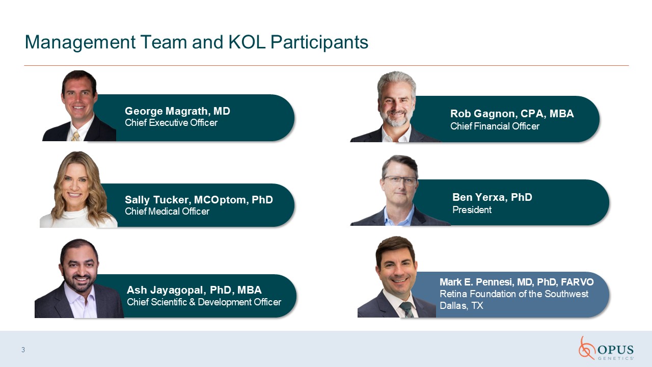
3 Management Team and KOL Participants Ash Jayagopal, PhD, MBA Chief Scientific
& Development Officer Sally Tucker, MCOptom, PhD Chief Medical Officer Rob Gagnon, CPA, MBA Chief Financial Officer Mark E. Pennesi, MD, PhD, FARVO Retina Foundation of the Southwest Dallas, TX George Magrath, MD Chief Executive
Officer Ben Yerxa, PhD President
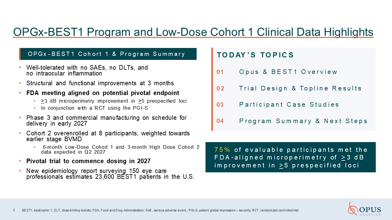
Well-tolerated with no SAEs, no DLTs, and no intraocular inflammation Structural
and functional improvements at 3 months FDA meeting aligned on potential pivotal endpoint >3 dB microperimetry improvement in >5 prespecified loci In conjunction with a RCT using the PGI-S Phase 3 and commercial manufacturing on
schedule for delivery in early 2027 Cohort 2 overenrolled at 8 participants, weighted towards earlier stage BVMD 6-month Low-Dose Cohort 1 and 3-month High Dose Cohort 2 data expected in Q2 2027 Pivotal trial to commence dosing in
2027 New epidemiology report surveying 150 eye care professionals estimates 23,600 BEST1 patients in the U.S. 4 BEST1, bestrophin 1; DLT, dose-limiting toxicity; FDA, Food and Drug Administration; SAE, serious adverse event.; PGI-S, patient
global impression – severity; RCT, randomized controlled trial OPGx-BEST1 Program and Low-Dose Cohort 1 Clinical Data Highlights OPGx - B E S T 1 C o h o r t 1 & P r o g r a m S u m m a r y T O D AY ’ S T O P I C S 01 O p u s &
B E S T 1 O v e r v i e w 0 2 T r i a l D e s i g n & To p l i n e R e s u l t s 03 P a r t i c i p a n t C a s e S t u d i e s 04 P r o g r a m S u m m a r y & N e x t S t e p s 7 5 % o f e v a l u a b l e p a r t i c i p a
n t s m e t t h e F D A - a l i g n e d m i c r o p e r i m e t r y of > 3 d B i m p r o v e m e n t i n > 5 p r e s p e c i f i e d l o c i
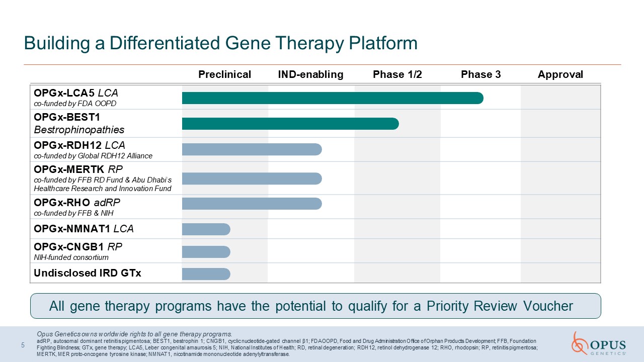
Building a Differentiated Gene Therapy Platform Opus Genetics owns worldwide
rights to all gene therapy programs. adRP, autosomal dominant retinitis pigmentosa; BEST1, bestrophin 1; CNGB1, cyclic nucleotide-gated channel β1; FDA OOPD, Food and Drug Administration Office of Orphan Products Development; FFB, Foundation
Fighting Blindness; GTx, gene therapy; LCA5, Leber congenital amaurosis 5; NIH, National Institutes of Health; RD, retinal degeneration; RDH12, retinol dehydrogenase 12; RHO, rhodopsin; RP, retinitis pigmentosa; MERTK, MER proto-oncogene
tyrosine kinase; NMNAT1, nicotinamide mononucleotide adenylyltransferase. 5 OPGx-LCA5 LCA co-funded by FDA OOPD OPGx-BEST1 Bestrophinopathies OPGx-RDH12 LCA co-funded by Global RDH12 Alliance OPGx-MERTK RP co-funded by FFB RD Fund
& Abu Dhabi’s Healthcare Research and Innovation Fund OPGx-RHO adRP co-funded by FFB & NIH OPGx-NMNAT1 LCA OPGx-CNGB1 RP NIH-funded consortium Undisclosed IRD GTx Preclinical IND-enabling Phase 1/2 Phase 3 Approval All gene
therapy programs have the potential to qualify for a Priority Review Voucher
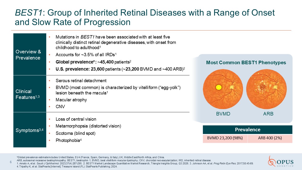
6 BEST1: Group of Inherited Retinal Diseases with a Range of Onset and Slow Rate
of Progression Overview & Prevalence Mutations in BEST1 have been associated with at least five clinically distinct retinal degenerative diseases, with onset from childhood to adulthood1 Accounts for ~3.5% of all IRDs1 Global
prevalence*: ~45,400 patients2 U.S. prevalence: 23,600 patients (~23,200 BVMD and ~400 ARB)2 Clinical Features1,3 Serous retinal detachment BVMD (most common) is characterized by vitelliform (“egg-yolk”) lesion beneath the
macula1 Macular atrophy CNV Symptoms3,4 Loss of central vision Metamorphopsia (distorted vision) Scotoma (blind spot) Photophobia4 *Global prevalence estimate includes United States, EU4 (France, Spain, Germany, & Italy), UK,
Middle East/North Africa, and China. ARB, autosomal recessive bestrophinopathy; BEST1, bestrophin 1; BVMD, best vitelliform macular dystrophy; CNV, choroidal neovascularization; IRD, inherited retinal disease. 1. Amato A, et al. Saudi J
Ophthalmol. 2023;37(4):287-295. 2. BEST1 Market Landscape Quantitative Market Research, Triangle Insights Group, Q3 2026. 3. Johnson AA, et al. Prog Retin Eye Res. 2017;58:45-69. 4. Tripathy K, et al. StatPearls [Internet]. Treasure Island
(FL): StatPearls Publishing; 2024. BVMD ARB Most Common BEST1 Phenotypes Prevalence BVMD 23,200 (98%) ARB 400 (2%)
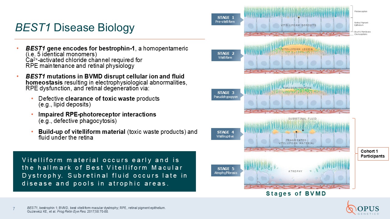
BEST1 gene encodes for bestrophin-1, a homopentameric (i.e. 5 identical
monomers) Ca2+-activated chloride channel required for RPE maintenance and retinal physiology BEST1 mutations in BVMD disrupt cellular ion and fluid homeostasis resulting in electrophysiological abnormalities, RPE dysfunction, and retinal
degeneration via: Defective clearance of toxic waste products (e.g., lipid deposits) Impaired RPE-photoreceptor interactions (e.g., defective phagocytosis) Build-up of vitelliform material (toxic waste products) and fluid under the
retina BEST1 Disease Biology BEST1, bestrophin 1; BVMD, best vitelliform macular dystrophy; RPE, retinal pigment epithelium. Guziewicz KE, et al. Prog Retin Eye Res. 2017;58:70-88. A T R O P H Y F R A G M E N T E D V I T E L L I F O R M
MA T E R I A L S U B R E T I N A L F L U I D P S E U D O H Y P O P Y O N Photoreceptors Retinal Pigment Epithelium Bruch’s Membrane Choriocapillaris V I T E L L I F O R M L E S I O N V I T E L L I F O R M D E P O S I T S STAGE
1 Pre-vitelliform STAGE 2 Vitelliform STAGE 3 Pseudohypopyon STAGE 4 Vitelliruptive STAGE 5 Atrophy/Fibrosis S t a g e s o f B V M D 7 Cohort 1 Participants V i t e l l i f o r m m a t e r i a l o c c u r s e a r l y a n d i s t
h e h a l l m a r k o f B e s t V i t e l l i f o r m M a c u l a r D y s t r o p h y. S u b r e t i n a l f l u i d o c c u r s l a t e i n d i s e a s e a n d p o o l s i n a t r o p h i c a r e a s .

8 Trial Design & Topline Cohort 1 Results Sally Tucker, MCOptom, PhD Chief
Medical Officer
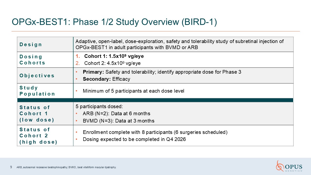
9 ARB, autosomal recessive bestrophinopathy; BVMD, best vitelliform macular
dystrophy. OPGx-BEST1: Phase 1/2 Study Overview (BIRD-1) D e s i g n Adaptive, open-label, dose-exploration, safety and tolerability study of subretinal injection of OPGx-BEST1 in adult participants with BVMD or ARB D o s i n g C o h o r
t s Cohort 1: 1.5x10⁹ vg/eye Cohort 2: 4.5x10⁹ vg/eye O b j e c t i v e s Primary: Safety and tolerability; identify appropriate dose for Phase 3 Secondary: Efficacy S t u d y P o p u l a t i o n Minimum of 5 participants at each dose
level S t a t u s o f C o h o r t 1 ( l o w d o s e ) 5 participants dosed: ARB (N=2): Data at 6 months BVMD (N=3): Data at 3 months S t a t u s o f C o h o r t 2 ( h i g h d o s e ) Enrollment complete with 8 participants (6
surgeries scheduled) Dosing expected to be completed in Q4 2026
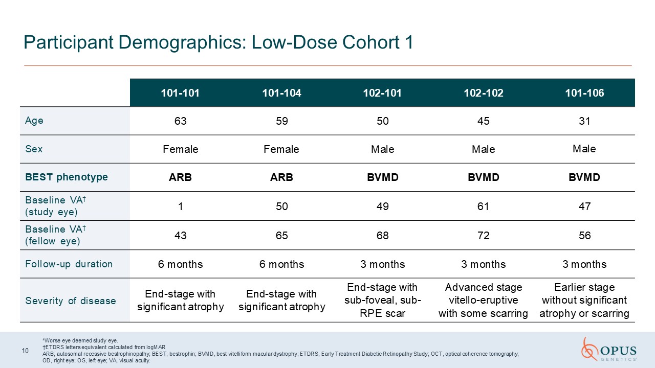
*Worse eye deemed study eye. †ETDRS letters equivalent calculated from
logMAR ARB, autosomal recessive bestrophinopathy; BEST, bestrophin; BVMD, best vitelliform macular dystrophy; ETDRS, Early Treatment Diabetic Retinopathy Study; OCT, optical coherence tomography; OD, right eye; OS, left eye; VA, visual
acuity. Participant Demographics: Low-Dose Cohort 1 101-101 101-104 102-101 102-102 101-106 Age 63 59 50 45 31 Sex Female Female Male Male Male BEST phenotype ARB ARB BVMD BVMD BVMD Baseline VA† (study
eye) 1 50 49 61 47 Baseline VA† (fellow eye) 43 65 68 72 56 Follow-up duration 6 months 6 months 3 months 3 months 3 months Severity of disease End-stage with significant atrophy End-stage with significant
atrophy End-stage with sub-foveal, sub-RPE scar Advanced stage vitello-eruptive with some scarring Earlier stage without significant atrophy or scarring 10
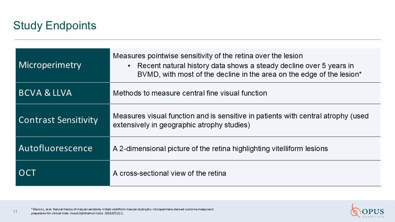
11 Study Endpoints Microperimetry Measures pointwise sensitivity of the retina
over the lesion Recent natural history data shows a steady decline over 5 years in BVMD, with most of the decline in the area on the edge of the lesion* BCVA & LLVA Methods to measure central fine visual function Contrast
Sensitivity Measures visual function and is sensitive in patients with central atrophy (used extensively in geographic atrophy studies) Autofluorescence A 2-dimensional picture of the retina highlighting vitelliform lesions OCT A
cross-sectional view of the retina * Bianco L, et al. Natural history of macular sensitivity in Best vitelliform macular dystrophy: microperimetry-derived outcome measures in preparation for clinical trials. Invest Ophthalmol Vis Sci.
2026;67(11):1.
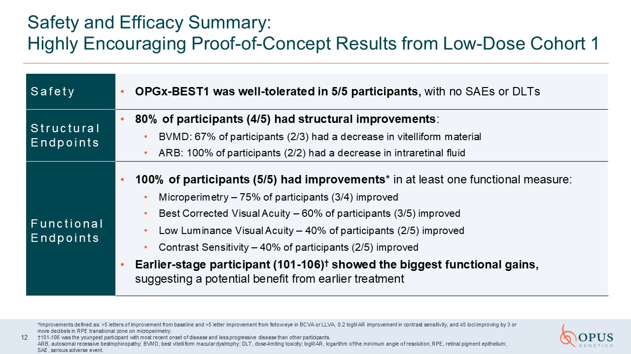
Safety and Efficacy Summary: Highly Encouraging Proof-of-Concept Results from
Low-Dose Cohort 1 S a f e t y OPGx-BEST1 was well-tolerated in 5/5 participants, with no SAEs or DLTs S t r u c t u r a l E n d p o i n t s 80% of participants (4/5) had structural improvements: BVMD: 67% of participants (2/3) had a
decrease in vitelliform material ARB: 100% of participants (2/2) had a decrease in intraretinal fluid F u n c t i o n a l E n d p o i n t s 100% of participants (5/5) had improvements* in at least one functional measure: Microperimetry –
75% of participants (3/4) improved Best Corrected Visual Acuity – 60% of participants (3/5) improved Low Luminance Visual Acuity – 40% of participants (2/5) improved Contrast Sensitivity – 40% of participants (2/5) improved Earlier-stage
participant (101-106)† showed the biggest functional gains, suggesting a potential benefit from earlier treatment *Improvements defined as: >5 letters of improvement from baseline and >5 letter improvement from fellow eye in BCVA or
LLVA, 0.2 logMAR improvement in contrast sensitivity, and ≥5 loci improving by 3 or more decibels in RPE transitional zone on microperimetry. †101-106 was the youngest participant with most recent onset of disease and less progressive
disease than other participants. ARB, autosomal recessive bestrophinopathy; BVMD, best vitelliform macular dystrophy; DLT, dose-limiting toxicity; logMAR, logarithm of the minimum angle of resolution; RPE, retinal pigment epithelium; SAE,
serious adverse event. 12
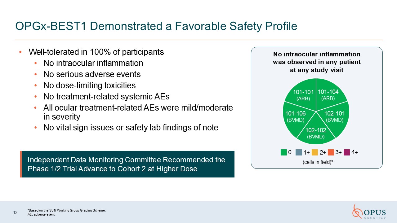
Well-tolerated in 100% of participants No intraocular inflammation No serious
adverse events No dose-limiting toxicities No treatment-related systemic AEs All ocular treatment-related AEs were mild/moderate in severity No vital sign issues or safety lab findings of note 13 OPGx-BEST1 Demonstrated a Favorable
Safety Profile Independent Data Monitoring Committee Recommended the Phase 1/2 Trial Advance to Cohort 2 at Higher Dose No intraocular inflammation was observed in any patient at any study visit *Based on the SUN Working Group Grading
Scheme. AE, adverse event. 101-101 101-104 (ARB) (ARB) 102-101 (BVMD) 101-106 (BVMD) 0 102-102 (BVMD) 1+ 2+ (cells in field)* 3+ 4+
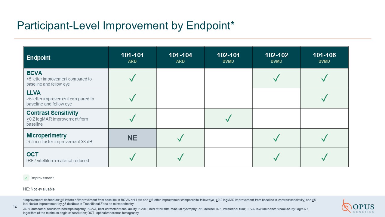
Participant-Level Improvement by
Endpoint* Endpoint 101-101 ARB 101-104 ARB 102-101 BVMD 102-102 BVMD 101-106 BVMD BCVA >5 letter improvement compared to baseline and fellow eye ✓ ✓ ✓ LLVA >5 letter improvement compared to baseline and fellow
eye ✓ ✓ Contrast Sensitivity >0.2 logMAR improvement from baseline ✓ ✓ Microperimetry >5 loci cluster improvement ≥3 dB NE ✓ ✓ ✓ OCT IRF / vitelliform material reduced ✓ ✓ ✓ ✓ Improvement NE: Not
evaluable *Improvement defined as: >5 letters of improvement from baseline in BCVA or LLVA and >5 letter improvement compared to fellow eye, >0.2 logMAR improvement from baseline in contrast sensitivity, and >5 loci cluster
improvement by >3 decibels in Transitional Zone on microperimetry. ARB, autosomal recessive bestrophinopathy; BCVA, best corrected visual acuity; BVMD, best vitelliform macular dystrophy; dB, decibel; IRF, intraretinal fluid; LLVA, low
luminance visual acuity; logMAR, logarithm of the minimum angle of resolution; OCT, optical coherence tomography. 14
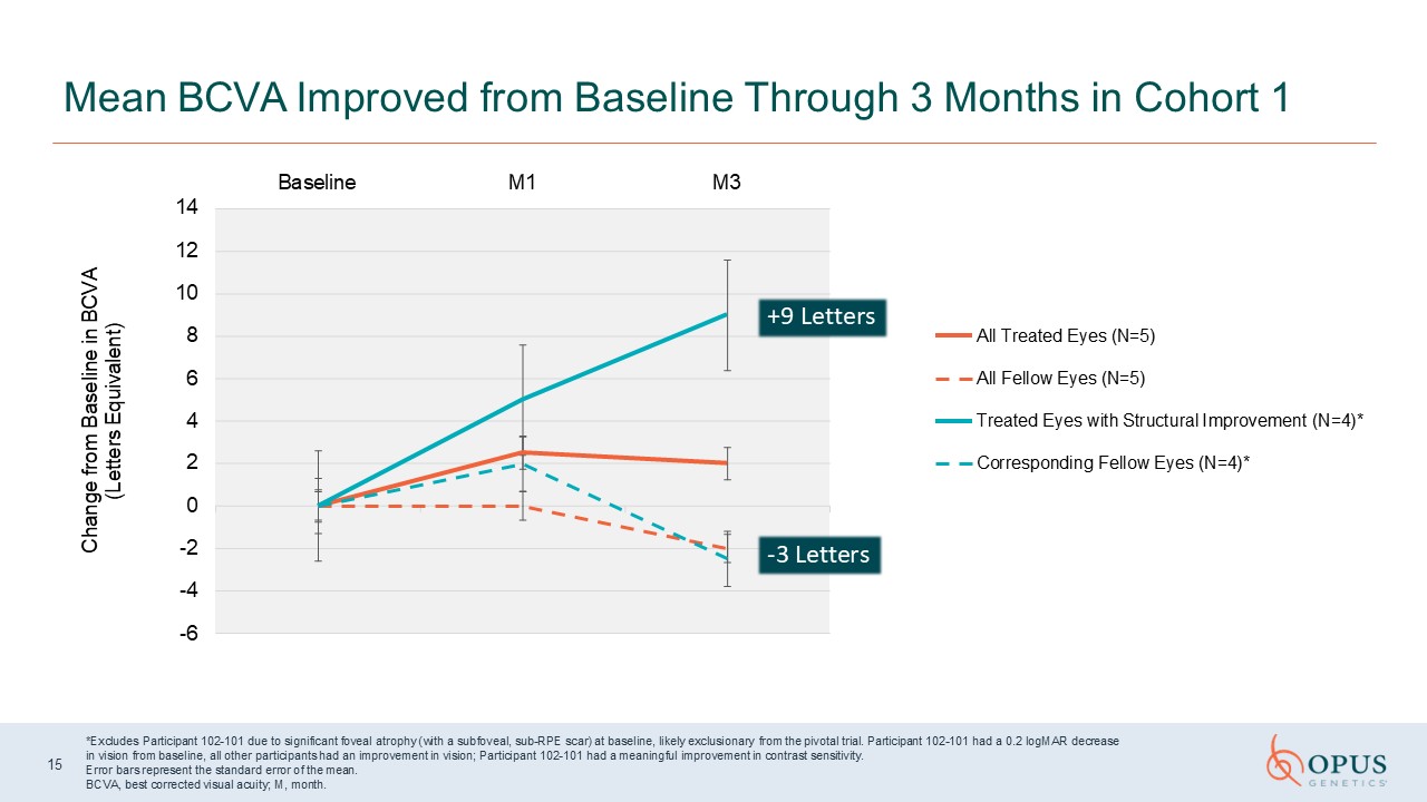
2 0 -2 -4 -6 14 12 10 8 6 4 Baseline M1 M3 Change from Baseline in
BCVA (Letters Equivalent) All Treated Eyes (N=5) All Fellow Eyes (N=5) Treated Eyes with Structural Improvement (N=4)* Corresponding Fellow Eyes (N=4)* Mean BCVA Improved from Baseline Through 3 Months in Cohort 1 *Excludes Participant
102-101 due to significant foveal atrophy (with a subfoveal, sub-RPE scar) at baseline, likely exclusionary from the pivotal trial. Participant 102-101 had a 0.2 logMAR decrease in vision from baseline, all other participants had an
improvement in vision; Participant 102-101 had a meaningful improvement in contrast sensitivity. Error bars represent the standard error of the mean. BCVA, best corrected visual acuity; M, month. 15 +9 Letters -3 Let ters
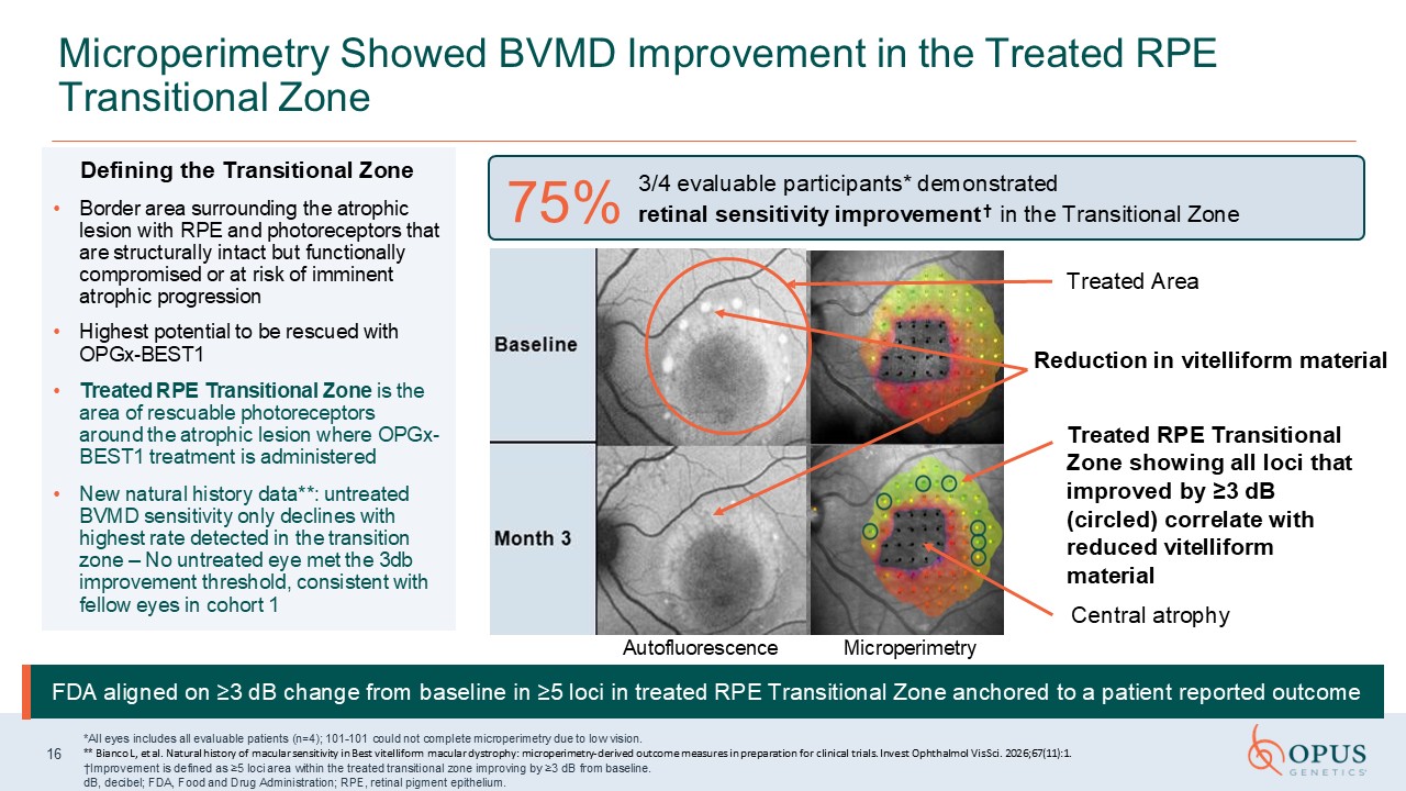
Defining the Transitional Zone Border area surrounding the atrophic lesion with
RPE and photoreceptors that are structurally intact but functionally compromised or at risk of imminent atrophic progression Highest potential to be rescued with OPGx-BEST1 Treated RPE Transitional Zone is the area of rescuable
photoreceptors around the atrophic lesion where OPGx-BEST1 treatment is administered New natural history data**: untreated BVMD sensitivity only declines with highest rate detected in the transition zone – No untreated eye met the 3db
improvement threshold, consistent with fellow eyes in cohort 1 Microperimetry Showed BVMD Improvement in the Treated RPE Transitional Zone 16 *All eyes includes all evaluable patients (n=4); 101-101 could not complete microperimetry due to
low vision. ** Bianco L, et al. Natural history of macular sensitivity in Best vitelliform macular dystrophy: microperimetry-derived outcome measures in preparation for clinical trials. Invest Ophthalmol Vis Sci. 2026;67(11):1. †Improvement
is defined as ≥5 loci area within the treated transitional zone improving by ≥3 dB from baseline. dB, decibel; FDA, Food and Drug Administration; RPE, retinal pigment epithelium. 3/4 evaluable participants* demonstrated † retinal
sensitivity improvement in the Transitional Zone 75% Treated Area Treated RPE Transitional Zone showing all loci that improved by ≥3 dB (circled) correlate with reduced vitelliform material Central atrophy Autofluorescence
Microperimetry FDA aligned on ≥3 dB change from baseline in ≥5 loci in treated RPE Transitional Zone anchored to a patient reported outcome Reduction in vitelliform material
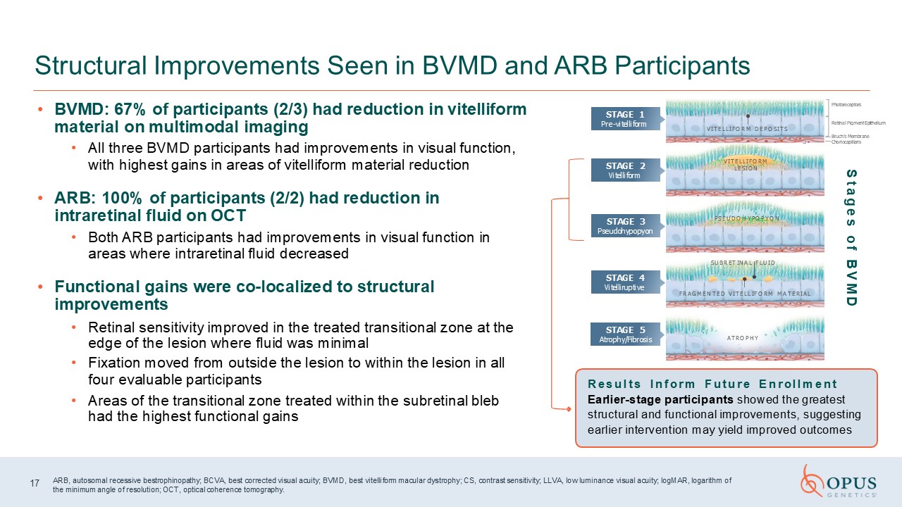
BVMD: 67% of participants (2/3) had reduction in vitelliform material on
multimodal imaging All three BVMD participants had improvements in visual function, with highest gains in areas of vitelliform material reduction ARB: 100% of participants (2/2) had reduction in intraretinal fluid on OCT Both ARB
participants had improvements in visual function in areas where intraretinal fluid decreased Functional gains were co-localized to structural improvements Retinal sensitivity improved in the treated transitional zone at the edge of the
lesion where fluid was minimal Fixation moved from outside the lesion to within the lesion in all four evaluable participants Areas of the transitional zone treated within the subretinal bleb had the highest functional gains Structural
Improvements Seen in BVMD and ARB Participants Results I nform Future E nrollment Earlier-stage participants showed the greatest structural and functional improvements, suggesting earlier intervention may yield improved outcomes ARB,
autosomal recessive bestrophinopathy; BCVA, best corrected visual acuity; BVMD, best vitelliform macular dystrophy; CS, contrast sensitivity; LLVA, low luminance visual acuity; logMAR, logarithm of the minimum angle of resolution; OCT,
optical coherence tomography. A T R O P H Y F R A G M E N T E D V I T E L L I F O R M M A T E R I A L S U B R E T I N A L F L U I D P S E U D O H Y P O P Y O N Photoreceptors Retinal Pigment Epithelium Bruch’s
Membrane Choriocapillaris V I T E L L I F O R M L E S I O N V I T E L L I F O R M D E P O S I T S STAGE 1 Pre-vitelliform STAGE 2 Vitelliform STAGE 3 Pseudohypopyon STAGE 4 Vitelliruptive STAGE 5 Atrophy/Fibrosis S t a g e s o f
B V M D 17
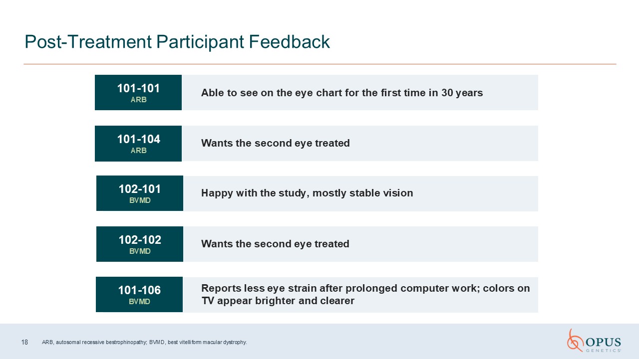
Post-Treatment Participant Feedback 101-101 ARB Able to see on the eye chart
for the first time in 30 years Wants the second eye treated 102-102 BVMD Wants the second eye treated Reports less eye strain after prolonged computer work; colors on TV appear brighter and clearer Happy with the study, mostly stable
vision 101-104 ARB 102-101 BVMD 101-106 BVMD 18 ARB, autosomal recessive bestrophinopathy; BVMD, best vitelliform macular dystrophy.

Participant Case Studies & Next Steps Ash Jayagopal, PhD Chief Scientific
and Development Officer 19
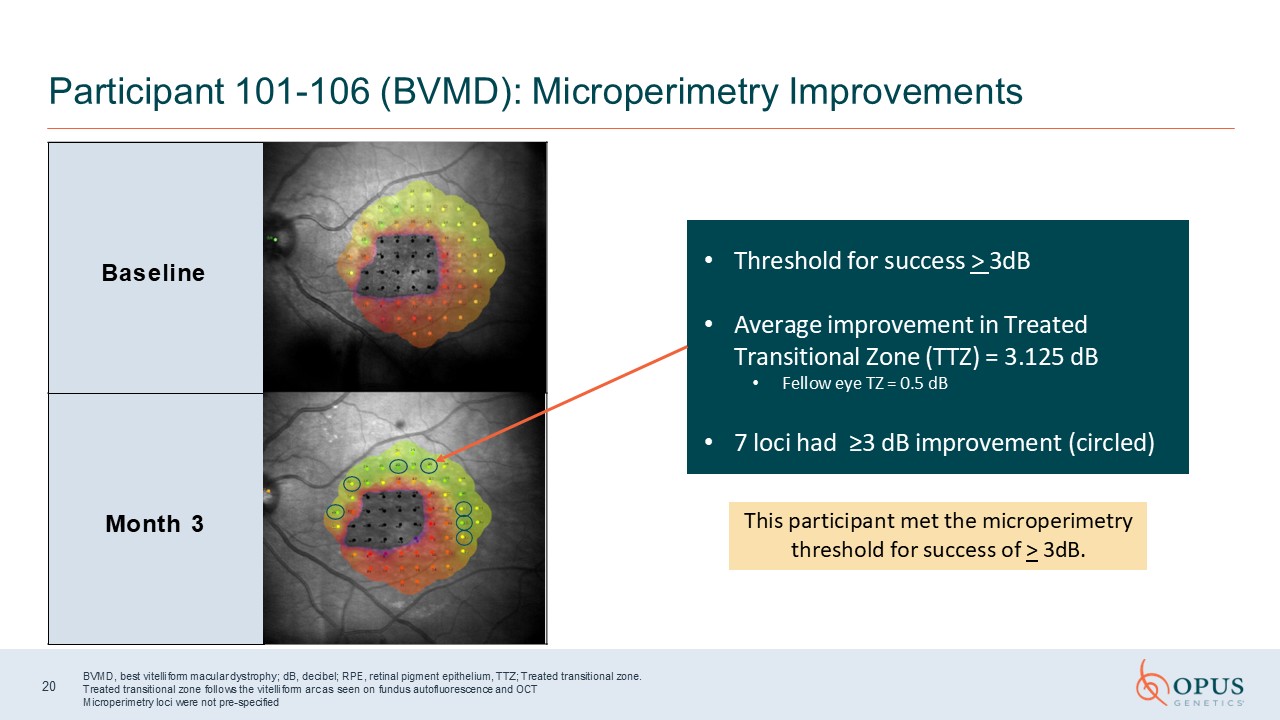
Baseline Month 3 Participant 101-106 (BVMD): Microperimetry
Improvements 20 BVMD, best vitelliform macular dystrophy; dB, decibel; RPE, retinal pigment epithelium, TTZ; Treated transitional zone. Treated transitional zone follows the vitelliform arc as seen on fundus autofluorescence and
OCT Microperimetry loci were not pre-specified Threshold for success > 3dB Average improvement in Treated Transitional Zone (TTZ) = 3.125 dB Fellow eye TZ = 0.5 dB 7 loci had ≥3 dB improvement (circled) This participant met the
microperimetry threshold for success of > 3dB.
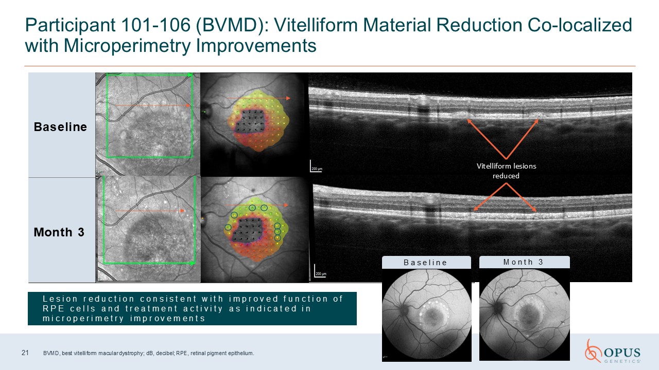
Baseline Month 3 Participant 101-106 (BVMD): Vitelliform Material Reduction
Co-localized with Microperimetry Improvements 21 BVMD, best vitelliform macular dystrophy; dB, decibel; RPE, retinal pigment epithelium. L e s i o n r e d u c t i o n c o n s i s t e n t w i t h i m p r o v e d f u n c t i o n o f R P E c
e l l s a n d t r e a t m e n t a c t i v i t y a s i n d i c a t e d i n m i c r o p e r i m e t r y i m p r o v e m e n t s Vitelliform lesions reduced B a s e l i n e M o n t h 3
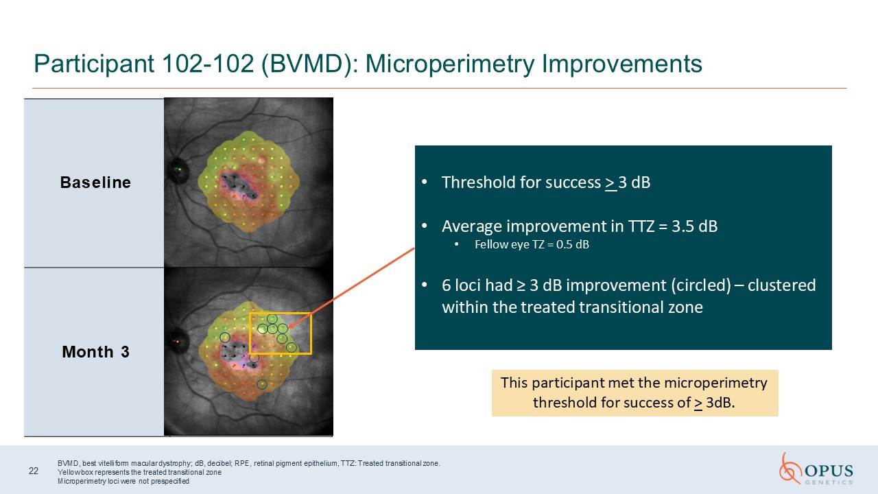
Baseline Month 3 Participant 102-102 (BVMD): Microperimetry
Improvements 22 BVMD, best vitelliform macular dystrophy; dB, decibel; RPE, retinal pigment epithelium, TTZ: Treated transitional zone. Yellow box represents the treated transitional zone Microperimetry loci were not
prespecified Threshold for success > 3 dB Average improvement in TTZ = 3.5 dB Fellow eye TZ = 0.5 dB 6 loci had ≥ 3 dB improvement (circled) – clustered within the treated transitional zone This participant met the microperimetry
threshold for success of > 3dB.
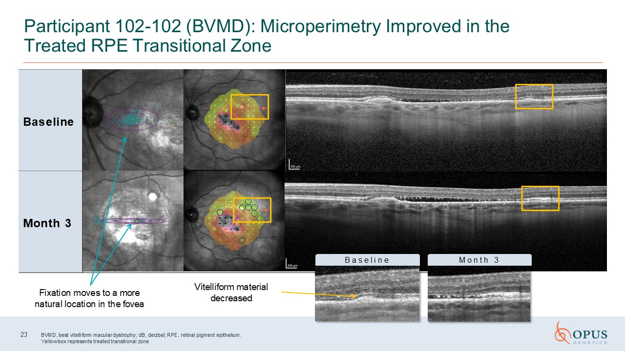
Baseline Month 3 Participant 102-102 (BVMD): Microperimetry Improved in the
Treated RPE Transitional Zone Baseline Month 3 B a s e l i n e M o n t h 3 23 Vitelliform material decreased BVMD, best vitelliform macular dystrophy; dB, decibel; RPE, retinal pigment epithelium. Yellow box represents treated
transitional zone Fixation moves to a more natural location in the fovea
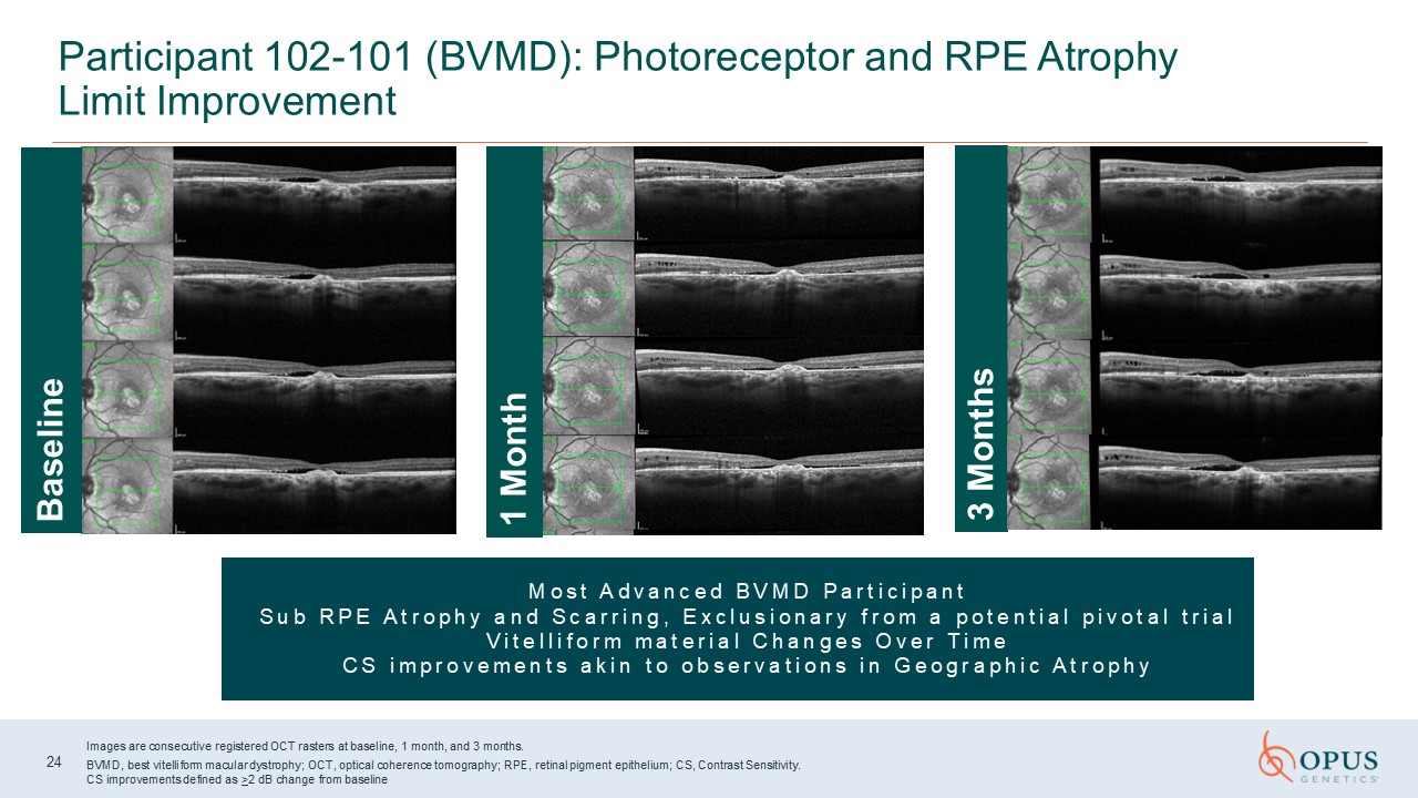
Participant 102-101 (BVMD): Photoreceptor and RPE Atrophy Limit
Improvement 24 Images are consecutive registered OCT rasters at baseline, 1 month, and 3 months. BVMD, best vitelliform macular dystrophy; OCT, optical coherence tomography; RPE, retinal pigment epithelium; CS, Contrast Sensitivity. CS
improvements defined as >2 dB change from baseline M o s t A d v a n c e d B V M D P a r t i c i p a n t S u b R P E A t r o p h y a n d S c a r r i n g , E x c l u s i o n a r y f r o m a p o t e n t i a l p i v o t a l t r i a l V i t
e l l i f o r m m a t e r i a l C h a n g e s O v e r T i m e C S i m p r o v e m e n t s a k i n t o o b s e r v a t i o n s i n G e o g r a p h i c A t r o p h y 3 Months Baseline 1 Month
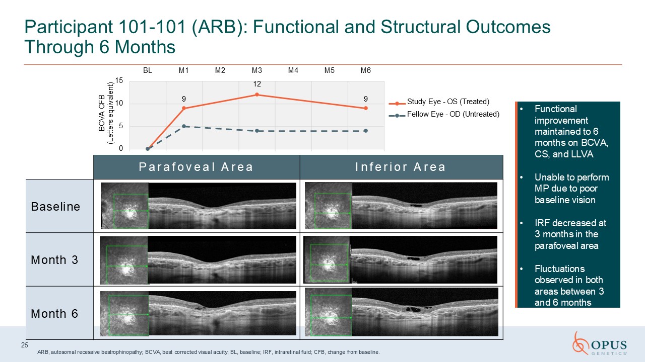
P a r a f o v e a l A r e a I n f e r i o r A r e a Baseline Month 3 Month
6 Participant 101-101 (ARB): Functional and Structural Outcomes Through 6 Months Functional improvement maintained to 6 months on BCVA, CS, and LLVA Unable to perform MP due to poor baseline vision IRF decreased at 3 months in the
parafoveal area Fluctuations observed in both areas between 3 and 6 months 25 ARB, autosomal recessive bestrophinopathy; BCVA, best corrected visual acuity; BL, baseline; IRF, intraretinal fluid; CFB, change from baseline. BL M1 M2 M3 M4
M5 M6 12 9 9 0 5 10 15 BCVA CFB (Letters equivalent) Study Eye - OS (Treated) Fellow Eye - OD (Untreated)
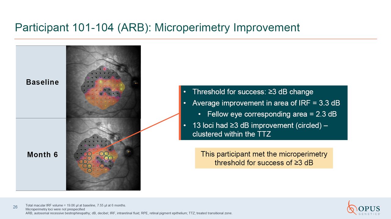
Participant 101-104 (ARB): Microperimetry
Improvement Baseline Month 6 26 Total macular IRF volume = 19.06 ul at baseline, 7.55 ul at 6 months. Microperimetry loci were not prespecified ARB, autosomal recessive bestrophinopathy; dB, decibel; IRF, intraretinal fluid; RPE, retinal
pigment epithelium. Threshold for success: > 3dB change Average improvement in area of IRF = 3.3 dB Fellow eye corresponding area = 2.3 dB 13 loci had ≥ 3 dB improvement (circled) – clustered within the treated transitional zone This
participant met the microperimetry threshold for success of > 3dB.
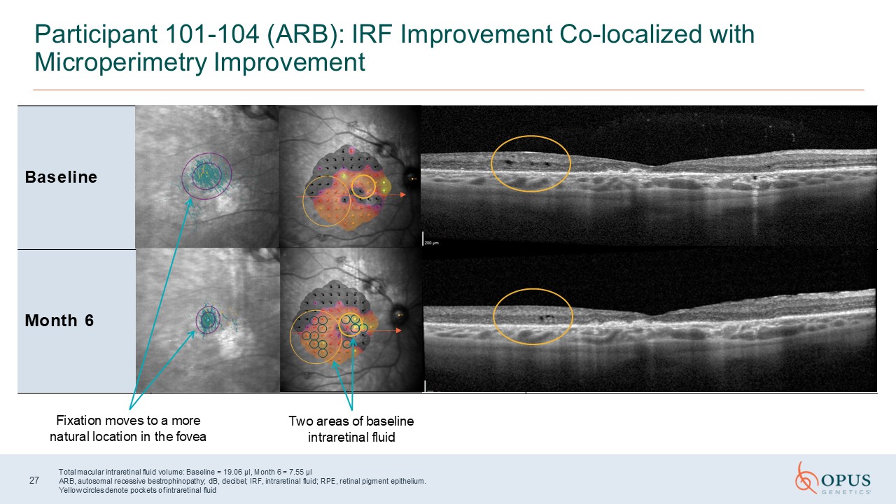
Participant 101-104 (ARB): IRF Improvement Co-localized with Microperimetry
Improvement Baseline Month 6 27 Total macular intraretinal fluid volume: Baseline = 19.06 µl, Month 6 = 7.55 µl ARB, autosomal recessive bestrophinopathy; dB, decibel; IRF, intraretinal fluid; RPE, retinal pigment epithelium. Yellow
circles denote pockets of intraretinal fluid Fixation moves to a more natural location in the fovea Two areas of baseline intraretinal fluid
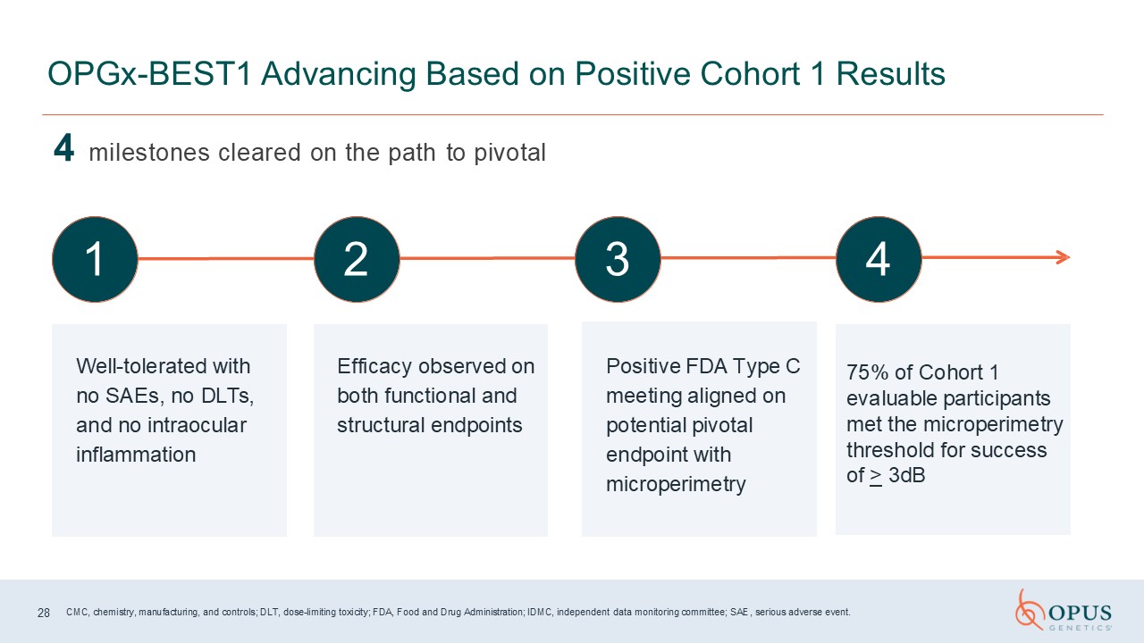
4 milestones cleared on the path to pivotal OPGx-BEST1 Advancing Based on
Positive Cohort 1 Results 1 2 3 4 Well-tolerated with no SAEs, no DLTs, and no intraocular inflammation Efficacy observed on both functional and structural endpoints Positive FDA Type C meeting aligned on potential pivotal endpoint with
microperimetry CMC, chemistry, manufacturing, and controls; DLT, dose-limiting toxicity; FDA, Food and Drug Administration; IDMC, independent data monitoring committee; SAE, serious adverse event. 28 75% of Cohort 1 evaluable participants
met the microperimetry threshold for success of > 3dB
Program Summary and Next Steps George Magrath, MD Chief Executive Officer 29
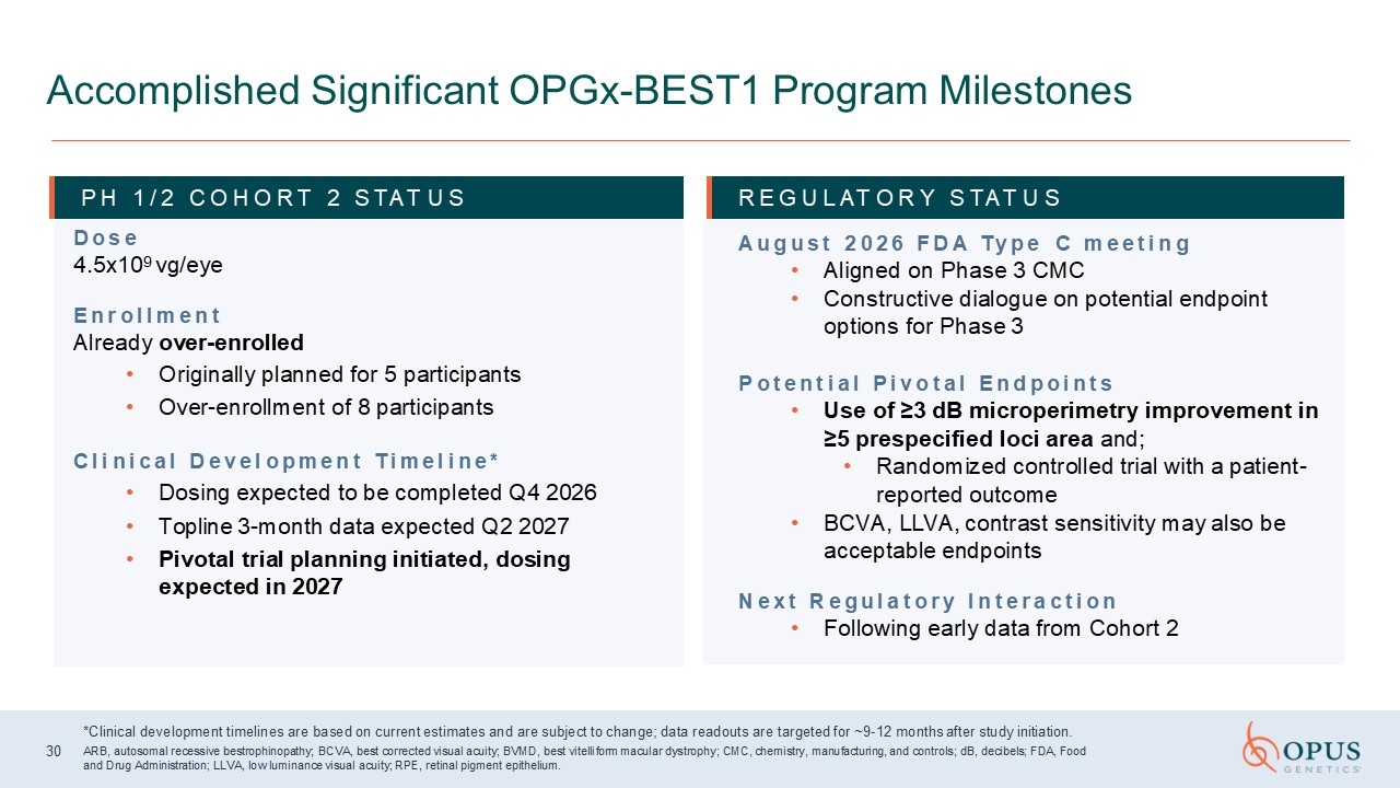
Accomplished Significant OPGx-BEST1 Program Milestones P H 1 / 2 C O H O R T 2 S
TAT U S D o s e 4.5x10⁹ vg/eye E n r o l l m e n t Already over-enrolled Originally planned for 5 participants Over-enrollment of 8 participants C l i n i c a l D e v e l o p m e n t T i m e l i n e * Dosing expected to be completed
Q4 2026 Topline 3-month data expected Q2 2027 Pivotal trial planning initiated, dosing expected in 2027 R E G U L AT O R Y S TAT U S A u g u s t 2 0 2 6 F D A T y p e C m e e t i n g Aligned on Phase 3 CMC Constructive dialogue on
potential endpoint options for Phase 3 P o t e n t i a l P i v o t a l E n d p o i n t s Use of ≥3 dB microperimetry improvement in ≥5 prespecified loci area and; Randomized controlled trial with a patient-reported outcome BCVA, LLVA,
contrast sensitivity may also be acceptable endpoints N e x t R e g u l a t o r y I n t e r a c t i o n Following early data from Cohort 2 *Clinical development timelines are based on current estimates and are subject to change; data
readouts are targeted for ~9-12 months after study initiation. ARB, autosomal recessive bestrophinopathy; BCVA, best corrected visual acuity; BVMD, best vitelliform macular dystrophy; CMC, chemistry, manufacturing, and controls; dB,
decibels; FDA, Food and Drug Administration; LLVA, low luminance visual acuity; RPE, retinal pigment epithelium. 30
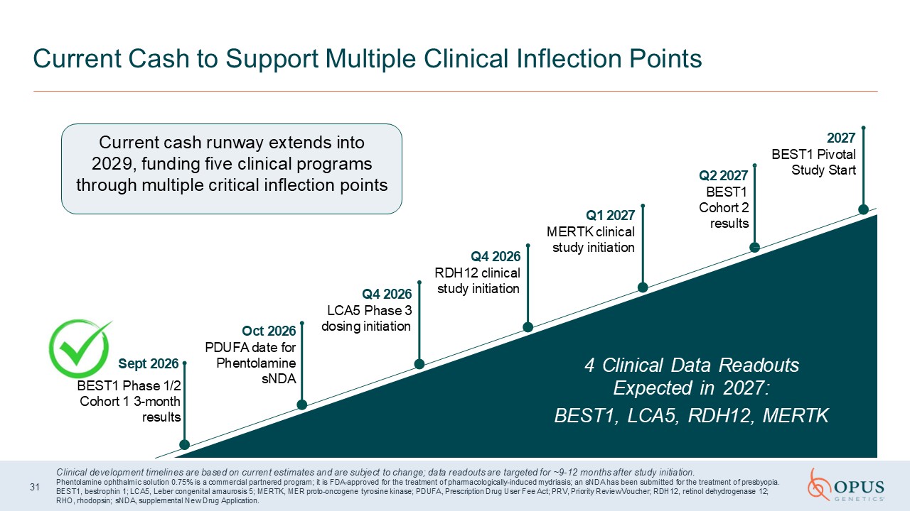
Current Cash to Support Multiple Clinical Inflection Points 2027 BEST1 Pivotal
Study Start Q1 2027 MERTK clinical study initiation Q4 2026 RDH12 clinical study initiation Q4 2026 LCA5 Phase 3 dosing initiation Oct 2026 PDUFA date for Phentolamine sNDA Sept 2026 BEST1 Phase 1/2 Cohort 1
3-month results Current cash runway extends into 2029, funding five clinical programs through multiple critical inflection points 4 Clinical Data Readouts Expected in 2027: BEST1, LCA5, RDH12, MERTK Clinical development timelines are
based on current estimates and are subject to change; data readouts are targeted for ~9-12 months after study initiation. Phentolamine ophthalmic solution 0.75% is a commercial partnered program; it is FDA-approved for the treatment of
pharmacologically-induced mydriasis; an sNDA has been submitted for the treatment of presbyopia. BEST1, bestrophin 1; LCA5, Leber congenital amaurosis 5; MERTK, MER proto-oncogene tyrosine kinase; PDUFA, Prescription Drug User Fee Act; PRV,
Priority Review Voucher; RDH12, retinol dehydrogenase 12; RHO, rhodopsin; sNDA, supplemental New Drug Application. Q2 2027 BEST1 Cohort 2 results 31

N a s d a q : I R D
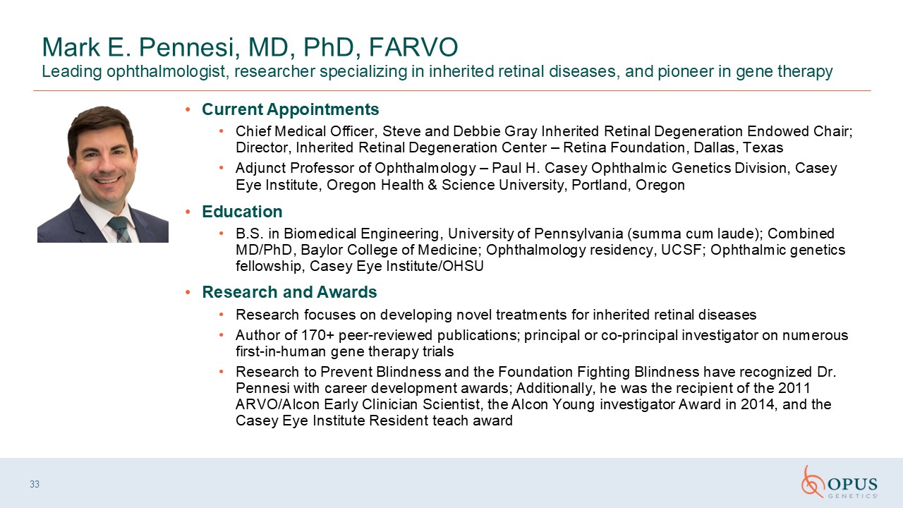
33 Mark E. Pennesi, MD, PhD, FARVO Leading ophthalmologist, researcher
specializing in inherited retinal diseases, and pioneer in gene therapy Current Appointments Chief Medical Officer, Steve and Debbie Gray Inherited Retinal Degeneration Endowed Chair; Director, Inherited Retinal Degeneration Center – Retina
Foundation, Dallas, Texas Adjunct Professor of Ophthalmology – Paul H. Casey Ophthalmic Genetics Division, Casey Eye Institute, Oregon Health & Science University, Portland, Oregon Education B.S. in Biomedical Engineering, University
of Pennsylvania (summa cum laude); Combined MD/PhD, Baylor College of Medicine; Ophthalmology residency, UCSF; Ophthalmic genetics fellowship, Casey Eye Institute/OHSU Research and Awards Research focuses on developing novel treatments for
inherited retinal diseases Author of 170+ peer-reviewed publications; principal or co-principal investigator on numerous first-in-human gene therapy trials Research to Prevent Blindness and the Foundation Fighting Blindness have recognized
Dr. Pennesi with career development awards; Additionally, he was the recipient of the 2011 ARVO/Alcon Early Clinician Scientist, the Alcon Young investigator Award in 2014, and the Casey Eye Institute Resident teach award
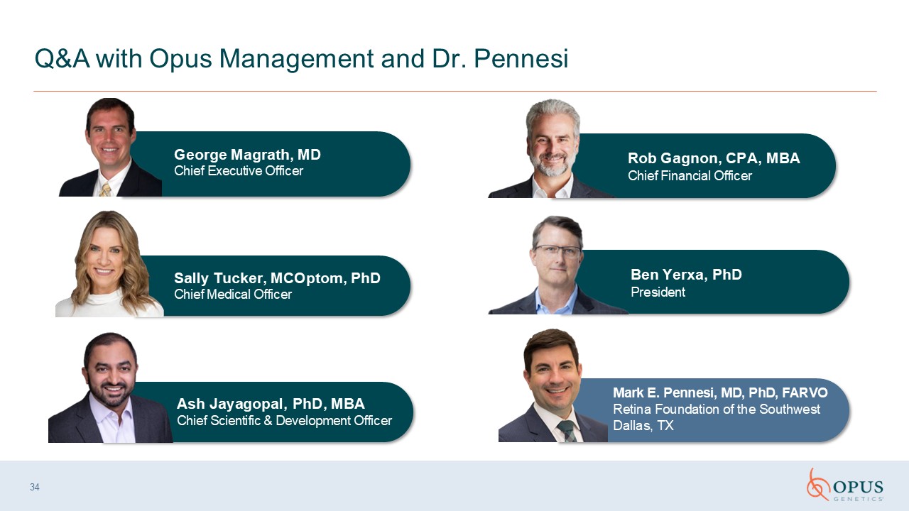
34 Q&A with Opus Management and Dr. Pennesi Ash Jayagopal, PhD, MBA Chief
Scientific & Development Officer Sally Tucker, MCOptom, PhD Chief Medical Officer Rob Gagnon, CPA, MBA Chief Financial Officer Mark E. Pennesi, MD, PhD, FARVO Retina Foundation of the Southwest Dallas, TX George Magrath, MD Chief
Executive Officer Ben Yerxa, PhD President
Appendix 35
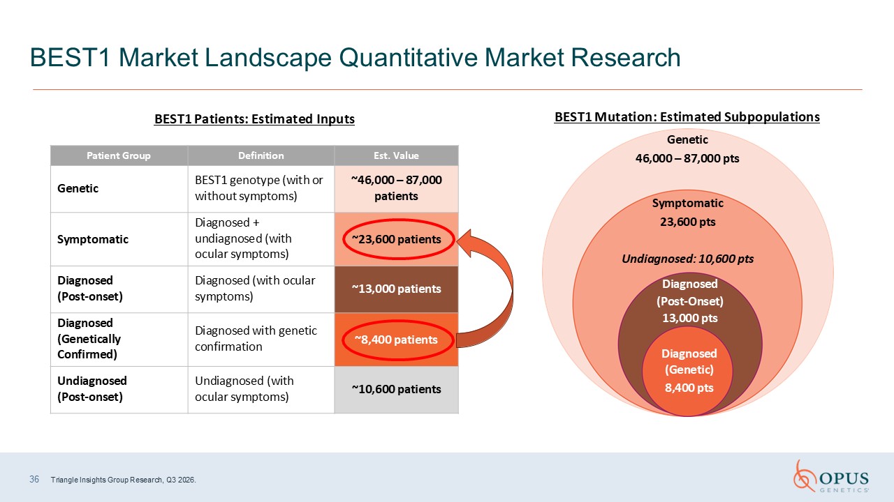
36 Triangle Insights Group Research, Q3 2026. BEST1 Market Landscape Quantitative
Market Research Patient Group Definition Est. Value Genetic BEST1 genotype (with or without symptoms) ~46,000 – 87,000 patients Symptomatic Diagnosed + undiagnosed (with ocular symptoms) ~23,600 patients Diagnosed
(Post-onset) Diagnosed (with ocular symptoms) ~13,000 patients Diagnosed (Genetically Confirmed) Diagnosed with genetic confirmation ~8,400 patients Undiagnosed (Post-onset) Undiagnosed (with ocular symptoms) ~10,600 patients BEST1
Patients: Estimated Inputs Symptomatic 23,600 pts Undiagnosed: 10,600 pts Diagnosed (Post-Onset) 13,000 pts Diagnosed (Genetic) 8,400 pts BEST1 Mutation: Estimated Subpopulations Genetic 46,000 – 87,000 pts